PE anti-mouse CD206 (MMR) Antibody

Pricing & Availability
Clone
C068C2 (See other available formats)
Regulatory Status
RUO
Other Names
MMR (macrophage mannose receptor), MR (mannose receptor), MRC1
Isotype
Rat IgG2a, κ
Ave. Rating
Submit a Review
Product Citations
publications
C068C2_PE_1_082911
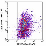
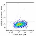
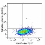
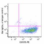
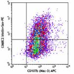
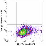
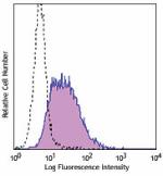
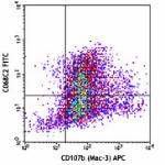
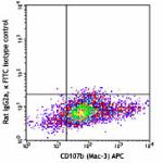
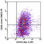
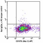
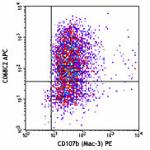
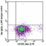
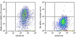
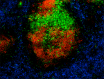
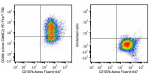
Thioglycollate-elicited BALB/c peritoneal macrophages were surface stained with CD107b (Mac-3) APC, and then intracellularly stained with CD206 (clone C068C2) PE (top) or rat IgG2a, κ PE isotype control (bottom).
  • C068C2_PE_1_082911
    Thioglycollate-elicited BALB/c peritoneal macrophages were surface stained with CD107b (Mac-3) APC, and then intracellularly stained with CD206 (clone C068C2) PE (top) or rat IgG2a, κ PE isotype control (bottom).
  • C068C2_PE_2_082911
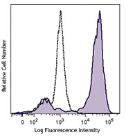
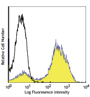
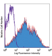
Compare all formats See PE spectral data
Cat # Size Price Quantity Check Availability Save
141705 25 µg 104€
Check Availability


Need larger quantities of this item?
Request Bulk Quote
141706 100 µg 240€
Check Availability


Need larger quantities of this item?
Request Bulk Quote
Description

CD206, also known as mannose receptor (MR), is a 175 kD type I membrane protein. It is a pattern recognition receptor (PRR) belonging to the C-type lectin superfamily. MR is expressed on macrophages, dendritic cells, Langerhans cells, and hepatic or lymphatic endothelial cells. MR recognizes a range of microbial carbohydrates bearing mannose, fucose, or N-acetyl glucosamine through its C-type lectin-like carbohydrate recognition domains, sulfated carbohydrate antigens through its cysteine-rich domain, and collagens through its fibronectin type II domain. MR mediates endocytosis and phagocytosis as well as activation of macrophages and antigen presentation. It plays an important role in host defense and provides a link between innate and adaptive immunity. Recently, MR on lymphatic endothelial cells was found to be involved in leukocyte trafficking and a contributor to the metastatic behavior of cancer cells. It suggests that MR may be a potential target in controlling inflammation and cancer metastasis by targeting the lymphatic vasculature.

Product Details
Technical Data Sheet (pdf)

Product Details

Verified Reactivity
Mouse
Antibody Type
Monoclonal
Host Species
Rat
Immunogen
Recombinant mouse CD206 (MMR)
Formulation
Phosphate-buffered solution, pH 7.2, containing 0.09% sodium azide.
Preparation
The antibody was purified by affinity chromatography and conjugated with PE under optimal conditions.
Concentration
0.2 mg/ml
Storage & Handling
The antibody solution should be stored undiluted between 2°C and 8°C, and protected from prolonged exposure to light. Do not freeze.
Application

ICFC - Quality tested
FC - Verified

Recommended Usage

Each lot of this antibody is quality control tested by intracellular immunofluorescent staining with flow cytometric analysis. For flow cytometric staining, the suggested use of this reagent is ≤0.5 µg per million cells in 100 µl volume.  It is recommended that the reagent be titrated for optimal performance for each application.

Excitation Laser
Blue Laser (488 nm)
Green Laser (532 nm)/Yellow-Green Laser (561 nm)
Application Notes

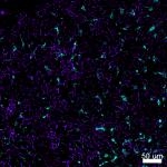
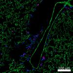
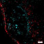
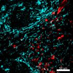
Clone C068C2 recognizes a region similar to clone MR5D3, based on the ability of the clones to block each other. Additional reported applications (for the relevant formats) include: spatial biology (IBEX)4,5.

Application References
  1. Keller J, et al. 2012. Biochem Biophys Res Commun. 417:217. PubMed
  2. Ito H, et al. 2012. J Am Soc Nephrol. 23:1797. PubMed
  3. Yang X, et al. 2015. PNAS. 112:2900. PubMed
  4. Radtke AJ, et al. 2020. Proc Natl Acad Sci U S A. 117:33455-65. (SB) PubMed
  5. Radtke AJ, et al. 2022. Nat Protoc. 17:378-401. (SB) PubMed
Product Citations
  1. Cui J, et al. 2021. J Cell Mol Med. 25:5586. PubMed
  2. Xiong Y, et al. 2020. J Nanobiotechnology. 18:66. PubMed
  3. Yu L, et al. 2021. Cell Biol Int. 45:2452. PubMed
  4. Li H, et al. 2022. Cancer Cell. 40:36. PubMed
  5. Silveira DB, et al. 2022. Cell Biochem Funct. 40:163. PubMed
  6. Liang X, et al. 2022. Cancer Commun (Lond). 42:205. PubMed
  7. Lee R, et al. 2022. Cancer Discov. 12:1702. PubMed
  8. Wang S, et al. 2022. Sci Adv. 8:eabn3883. PubMed
  9. Li D, et al. 2022. Cell Prolif. 55:e13299. PubMed
  10. Li Y, et al. 2023. Bioact Mater. 20:548. PubMed
  11. Liu Z, et al. 2022. Cancer Commun (Lond). 42:971. PubMed
  12. Meng Z, et al. 2022. Int J Oncol. 61: . PubMed
  13. Zhu J, et al. 2022. Nat Commun. 13:7466. PubMed
  14. Li J, et al. 2023. Front Immunol. 13:945234. PubMed
  15. Choi H, et al. 2023. J Nanobiotechnology. 21:5. PubMed
  16. Domingos-Pereira S, et al. 2022. Int J Mol Sci. 24: . PubMed
  17. Li Y, et al. 2023. Cell Death Dis. 14:28. PubMed
  18. Zhang L, et al. 2023. J Inflamm Res. 16:109. PubMed
  19. Fan R, et al. 2023. J Immunother Cancer. 11: . PubMed
  20. Chun J, et al. 2023. Cancers (Basel). 15: . PubMed
  21. Li S, et al. 2023. Exp Mol Med. 55:457. PubMed
  22. Zhao X, et al. 2022. Cell Mol Immunol. 19:540. PubMed
  23. Lundahl MLE, et al. 2022. Elife. 11:. PubMed
  24. Adzika GK, et al. 2023. Front Immunol. 14:1124649. PubMed
  25. Bautista CA, et al. 2023. Front Physiol. 14:1122348. PubMed
  26. Garabuczi É, et al. 2023. Front Immunol. 14:1139204. PubMed
  27. Chung JY, et al. 2023. Nat Commun. 14:1794. PubMed
  28. Shi Y, et al. 2023. Nat Commun. 14:1884. PubMed
  29. Pan C, et al. 2023. Front Bioeng Biotechnol. 11:1129187. PubMed
  30. Levra Levron C, et al. 2023. Nat Cell Biol. 25:740. PubMed
  31. Seclì L, et al. 2023. J Immunother Cancer. 11:. PubMed
  32. Li T, et al. 2023. Nat Commun. 14:2498. PubMed
  33. Salemme V, et al. 2023. Nat Commun. 14:2350. PubMed
  34. Zeng S, et al. 2023. Front Oncol. 13:1171926. PubMed
  35. Michela Miani et al. 2018. Cell metabolism. 28(4):557-572 . PubMed
  36. Feng Y, et al. 2018. J Am Soc Nephrol. 29:182. PubMed
  37. Seo JB, et al. 2019. Nat Metab. 1:86. PubMed
  38. Ying W, et al. 2019. Cell Metab. 29:457. PubMed
  39. Sun H, et al. 2019. Mol Hum Reprod. 25:05:00. PubMed
  40. Zhang B, et al. 2022. Front Med (Lausanne). 8:796743. PubMed
  41. Wang HH, et al. 2022. J Biomed Sci. 29:36. PubMed
  42. Zhang Z, et al. 2022. Front Pharmacol. 13:906625. PubMed
  43. Hu S, et al. 2018. Front Immunol. 9:01. PubMed
  44. Ying W et al. 2017. Cell. 171(2):372-384 . PubMed
  45. Tripathi H, et al. 2020. J Mol Cell Cardiol. 149:95. PubMed
  46. Leach SM, et al. 2020. Cell Rep. 33:108337. PubMed
  47. Zheng W, et al. 2021. Wellcome Open Research. 6:52. PubMed
  48. Uzhachenko RV, et al. 2021. Cell Reports. 35(1):108944. PubMed
  49. Dong L, et al. 2021. Cancer Cell. . PubMed
  50. Sadek J, et al. 2021. EMBO Mol Med. 13:e13591. PubMed
  51. Hsu SPC, et al. 2020. J Neurooncol. 146:417. PubMed
  52. Nomura M, et al. 2016. Nat Immunol. 17:216-217. PubMed
  53. Clarke F,et al. 2017. PLoS One. 10.1371/journal.pone.0186625. PubMed
  54. Zhang Y, et al. 2018. Sci Rep. 8:5994. PubMed
  55. Li Q, et al. 2021. Front Immunol. 12:707784. PubMed
  56. Pajarinen J, et al. 2021. J Biomed Mater Res A. 109:1512. PubMed
  57. Zheng H, et al. 2021. Frontiers in Cell and Developmental Biology. 9:641527. PubMed
  58. De Vlaeminck Y, et al. 2020. Cancers (Basel). 12:. PubMed
  59. Li J, et al. 2021. Front Immunol. 12:649285. PubMed
  60. Krukowski K, et al. 2021. Sci Adv. 7:eabg6702. PubMed
  61. Shao H, et al. 2012. J Biol Chem. 287:35860. PubMed
  62. Jia X, et al. 2021. Front Cell Dev Biol. 9:685522. PubMed
  63. He C, et al. 2021. Oncoimmunology. 1897295:10. PubMed
  64. Tang TT, et al. 2020. Sci Adv. 6:eaaz0748. PubMed
  65. Mul Fedele ML, et al. 2020. Front Cell Infect Microbiol. 10:100. PubMed
  66. El–Achkar GA, et al. 2019. PLoS One. 14:e0216405. PubMed
  67. Zhang J, et al. 2021. J Leukoc Biol. 110:485. PubMed
  68. Tanner L, et al. 2022. Front Pharmacol. 13:899469. PubMed
  69. Hou H, et al. 2021. Front Cell Dev Biol. 9:737003. PubMed
  70. Johansen AZ, et al. 2022. Pharmaceutics. 14:. PubMed
  71. McKee AM, et al. 2021. iScience. 24:103012. PubMed
  72. Giri J, et al. 2020. Cell Rep. 30:1923. PubMed
  73. Dan H, et al. 2020. Mol Oncol. 14:795. PubMed
  74. Kim JS, et al. 2020. Int J Oncol. 56:1405. PubMed
  75. Rao X, et al. 2022. Cell Death Dis. 13:891. PubMed
  76. Zhang Y, et al. 2022. Nat Commun. 13:4553. PubMed
  77. Martins L, et al. 2020. Stem Cell Res Ther. 0.786805556. PubMed
  78. Farini A, et al. 2020. Front Physiol. 0.738194444. PubMed
  79. An W et al. 2019. British journal of pharmacology. 176(5):699-710 . PubMed
  80. Li J, et al. 2021. Cell Commun Signal. 19:89. PubMed
  81. Mehta AK, et al. 2021. Nat Cancer. 2:66. PubMed
  82. Jiao Y, et al. 2021. Crit Care. 25:356. PubMed
  83. Liao T, et al. 2017. Front Immunol. 8:1334. PubMed
  84. Guérin MV, et al. 2019. Nat Commun. 10:4131. PubMed
  85. Wang J, et al. 2021. Nat Commun. 12:6198. PubMed
  86. Lu X, et al. 2020. Sci Transl Med. 12:. PubMed
  87. Cai C, et al. 2021. Sci Rep. 11:13386. PubMed
  88. Toubal A, et al. 2020. Nat Commun. 3755:11. PubMed
  89. Tacconi C, et al. 2021. Cell Reports. 35(2):108993. PubMed
  90. Wei W, et al. 2022. mSystems. 7:e0046922. PubMed
  91. Lee DH, et al. 2019. PLoS One. 14:e0215727. PubMed
  92. Yu Y, et al. 2018. Mol Med Rep. 17:8212. PubMed
  93. Wang Y, et al. 2022. Cell Death Dis. 13:748. PubMed
  94. Golden TN, et al. 2022. Front Pharmacol. 12:761496. PubMed
  95. Miura I, et al. 2021. Physiol Rep. 9:e14859. PubMed
  96. Ruiz de Azua I, et al. 2017. J Clin Invest. 127:4148. PubMed
  97. Li H, et al. 2021. Nat Commun. 12:7149. PubMed
  98. Germundson DL, et al. 2022. Front Allergy. 3:870513. PubMed
  99. Shu Y, et al. 2020. Immunology. 160:345. PubMed
  100. Sztwiertnia I, et al. 2020. PLoS One. 15:e0233789. PubMed
  101. Ying W, et al. 2016. Sci Rep. 6:20176. PubMed
  102. Liu C et al. 2019. Immunity. 51(2):381-397 . PubMed
  103. Chu SY, et al. 2019. Nat Commun. 10:1524. PubMed
  104. Anderson AE, et al. 2022. NPJ Regen Med. 7:6. PubMed
  105. Lu X, et al. 2012. Arterioscler Thromb Vasc Biol. 32:2358. PubMed
  106. Bhattacharya A, et al. 2018. Neuropsychopharmacology. 43:2586. PubMed
  107. Qiu N, et al. 2021. J Nanobiotechnology. 19:428. PubMed
  108. Yang C, et al. 2022. Nat Commun. 13:4866. PubMed
  109. Lowe PP, et al. 2020. J Neuroinflammation. 0.913888889. PubMed
  110. Lu SW, et al. 2020. Nat Commun. 3.660416667. PubMed
  111. Zhou K, et al. 2016. J Cereb Blood Flow Metab. 10.1177/0271678X16648712. PubMed
  112. Deng W, et al. 2015. Clin Immunol. . PubMed
  113. Anderson-Baucum E, et al. 2021. Cell Metab. 33:1883. PubMed
  114. Stevenson ER, et al. 2022. J Pharmacol Exp Ther. 382:356. PubMed
  115. Wang X, et al. 2022. J Biol Chem. 298:101561. PubMed
  116. Asterholm I, et al. 2012. J Lipid Res. 53: 1254. PubMed
  117. Stridh L, et al. 2013. J Neurosci. 33:12401. PubMed
  118. Zhu Q, et al. 2022. Atherosclerosis. 357:41. PubMed
  119. Long L, et al. 2018. Bio Protoc. 8:e2815. PubMed
  120. Li C, et al. 2019. JCI Insight. 5. PubMed
  121. Budai Z, et al. 2021. Cells. 10:. PubMed
  122. Suzuki T, et al. 2017. Cell Rep. 18(8):2045-2057. PubMed
  123. Krukowski K, et al. 2018. Sci Rep. 8:7857. PubMed
  124. Di Lorenzo A, et al. 2022. Oncoimmunology. 11:2086752. PubMed
  125. Li Y, et al. 2020. Theranostics. 10:2897. PubMed
  126. Leary N, et al. 2022. J Extracell Vesicles. 11:e12197. PubMed
  127. Zhang W, et al. 2021. Front Immunol. 12:768435. PubMed
  128. Santopaolo M, et al. 2021. Front Immunol. 609406:12. PubMed
  129. Sears SM, et al. 2022. Kidney360. 3:818. PubMed
  130. Ukah T, et al. 2017. J Immunol. 10.4049/jimmunol.1700410. PubMed
  131. Roediger B et al. 2018. Cell. 175(2):530-543 . PubMed
  132. Liu C, et al. 2022. Cell Death Discov. 8:269. PubMed
  133. Liu YS, et al. 2021. Cancers (Basel). 13:. PubMed
  134. Budhu S, et al. 2021. Cell Reports. 34(2):108620. PubMed
  135. Srivastava S, et al. 2020. Cancer Cell. 39(2):193-208.e10. PubMed
  136. Zhang J, et al. 2020. J Cell Mol Med. 24:4547. PubMed
  137. Browne A, et al. 2015. Cancer Res . 75: 2811 - 2821. PubMed
  138. Moreno M et al. 2014. The Journal of Neuroscience. 34(24):8175-85 . PubMed
  139. Lau P, et al. 2022. Cell Mol Immunol. :. PubMed
  140. Al-Zaeed N, et al. 2021. Cell Death Dis. 12:611. PubMed
  141. Golabek A, et al. 2021. Exp Ther Med. 21:482. PubMed
  142. Yuan Y, et al. 2019. Clin Sci (Lond). 133:1759. PubMed
  143. Hong Y, et al. 2019. J Extracell Vesicles. 8:1670893. PubMed
  144. Sadtler K, et al. 2017. Tissue Eng Part A. 23:1044. PubMed
  145. Li B et al. 2019. Cell reports. 26(10):2720-2737 . PubMed
  146. Morioka S, et al. 2018. Nature. 563:714. PubMed
  147. Mellal K, et al. 2019. Sci Rep. 9:12903. PubMed
  148. Yeini E, et al. 2021. Nat Commun. 12:1912. PubMed
  149. Xu GY, et al. 2022. Small. 18:e2107838. PubMed
  150. Zheng H, et al. 2021. Frontiers in Immunology. 12:645100. PubMed
  151. Cai T, et al. 2022. Front Immunol. 13:1009932. PubMed
  152. Chuan Li et al. 2017. Endocrinology. 158(9):2837-2847 . PubMed
  153. Wen HJ, et al. 2019. Cell Mol Gastroenterol Hepatol. 8:p173. PubMed
  154. Yuan M, et al. 2017. Hum Reprod. 32:94. PubMed
  155. Riopel M, et al. 2019. Mol Metab. 20:89. PubMed
  156. Aurélien Trompette et al. 2018. Immunity. 48(5):992-1005 . PubMed
  157. Lal JC, et al. 2021. Breast Cancer Res. 23:83. PubMed
  158. Guo S, et al. 2021. Chronic Dis Transl Med. 7:254. PubMed
RRID
AB_10896421 (BioLegend Cat. No. 141705)
AB_10895754 (BioLegend Cat. No. 141706)

Antigen Details

Structure
Type I transmembrane protein, 175 kD, C-type lectin superfamily
Distribution

Macrophages, dendritic cells, Langerhans cells, liver endothelial cells

Function
Pathogen recognition, endocytosis and phagocytosis, antigen presentation
Ligand/Receptor
Antigen containing mannose, fucose, or an N-acetyl glucosamine
Cell Type
Dendritic cells, Endothelial cells, Langerhans cells, Macrophages
Biology Area
Cell Biology, Immunology, Innate Immunity, Signal Transduction
Molecular Family
CD Molecules
Antigen References

1. Wileman TE, et al. 1986. P. Natl. Acad. Sci. USA 83:2501.
2. Apostolopoulos V, et al. 2001. Curr. Mol. Med. 1:469.
3. Burgdorf S, et al. 2006. J. Immunol. 176:6770.
4. McKenzie EJ, et al. 2007. J. Immunol. 178:4975.

Gene ID
17533 View all products for this Gene ID
UniProt
View information about CD206 on UniProt.org

Related FAQs

What type of PE do you use in your conjugates?
We use R-PE in our conjugates.
Why is mouse CD206 stained intracellularly and not via surface staining?

Typically, mouse CD206 surface level is relatively low under normal conditions and so intracellular staining protocol is required to get better signal.

Other Formats

View All CD206 Reagents Request Custom Conjugation
Description Clone Applications
Brilliant Violet 711™ anti-mouse CD206 (MMR) C068C2 ICFC,FC
Brilliant Violet 785™ anti-mouse CD206 (MMR) C068C2 ICFC,FC
PE/Dazzle™ 594 anti-mouse CD206 (MMR) C068C2 ICFC,FC
Alexa Fluor® 700 anti-mouse CD206 (MMR) C068C2 ICFC,FC
Spark YG™ 570 anti-mouse CD206 (MMR) C068C2 IHC-F
Biotin anti-mouse CD206 (MMR) C068C2 ICFC,FC
Purified anti-mouse CD206 (MMR) C068C2 ICFC,FC
FITC anti-mouse CD206 (MMR) C068C2 ICFC,FC
PE anti-mouse CD206 (MMR) C068C2 ICFC,FC
APC anti-mouse CD206 (MMR) C068C2 ICFC,FC
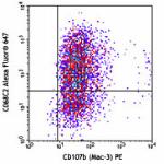
Alexa Fluor® 647 anti-mouse CD206 (MMR) C068C2 ICFC,FC,IHC-F
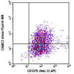
Alexa Fluor® 488 anti-mouse CD206 (MMR) C068C2 ICFC,FC,IHC-F
PerCP/Cyanine5.5 anti-mouse CD206 (MMR) C068C2 ICFC,FC
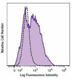
PE/Cyanine7 anti-mouse CD206 (MMR) C068C2 ICFC,FC
Brilliant Violet 421™ anti-mouse CD206 (MMR) C068C2 ICFC,FC,IHC-F,SB
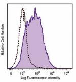
Brilliant Violet 605™ anti-mouse CD206 (MMR) C068C2 ICFC,FC
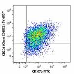
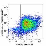
Brilliant Violet 650™ anti-mouse CD206 (MMR) C068C2 ICFC,FC
Alexa Fluor® 594 anti-mouse CD206 (MMR) C068C2 IHC-F
PE/Cyanine5 anti-mouse CD206 (MMR) C068C2 ICFC,FC
PE/Fire™ 700 anti-mouse CD206 (MMR) C068C2 ICFC,FC
TotalSeq™-B0173 anti-mouse CD206 (MMR) C068C2 ICPG
Spark Red™ 718 anti-mouse CD206 (MMR) (Flexi-Fluor™) C068C2 FC
Spark Blue™ 574 anti-mouse CD206 (MMR) (Flexi-Fluor™) C068C2 FC
TotalSeq™-C0173 anti-mouse CD206 (MMR) Antibody C068C2 ICPG
Spark YG™ 581 anti-mouse CD206 (MMR) (Flexi-Fluor™) C068C2 FC
Spark YG™ 593 anti-mouse CD206 (MMR) (Flexi-Fluor™) Antibody C068C2 FC
Spark NIR™ 685 anti-mouse CD206 (MMR) (Flexi-Fluor™) Antibody C068C2 FC
Go To Top Version: 3    Revision Date: 04/04/2016

For Research Use Only. Not for diagnostic or therapeutic use.


This product is supplied subject to the terms and conditions, including the limited license, located at www.biolegend.com/terms) ("Terms") and may be used only as provided in the Terms. Without limiting the foregoing, BioLegend products may not be used for any Commercial Purpose as defined in the Terms, resold in any form, used in manufacturing, or reverse engineered, sequenced, or otherwise studied or used to learn its design or composition without express written approval of BioLegend. Regardless of the information given in this document, user is solely responsible for determining any license requirements necessary for user’s intended use and assumes all risk and liability arising from use of the product. BioLegend is not responsible for patent infringement or any other risks or liabilities whatsoever resulting from the use of its products.


BioLegend, the BioLegend logo, and all other trademarks are property of BioLegend, Inc. or their respective owners, and all rights are reserved.


8999 BioLegend Way, San Diego, CA 92121 www.biolegend.com
Toll-Free Phone: 1-877-Bio-Legend (246-5343) Phone: (858) 768-5800 Fax: (877) 455-9587

This data display is provided for general comparisons between formats.
Your actual data may vary due to variations in samples, target cells, instruments and their settings, staining conditions, and other factors.
If you need assistance with selecting the best format contact our expert technical support team.

ProductsHere

Login / Register
Remember me
Forgot your password? Reset password?
Create an Account