


- Clone
- TC15-12F12.2 (See other available formats)
- Regulatory Status
- RUO
- Other Names
- Signaling Lymphocyte Activation Molecule (SLAM), IPO-3
- Isotype
- Rat IgG2a, λ
- Ave. Rating
- Submit a Review
- Product Citations
- publications

-

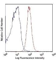
C57BL/6 mouse splenocytes were stained with CD150 (clone TC15-12F12.2) PE (solid line) or rat IgG2a PE isotype control (broken line). -

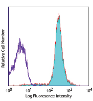
C57BL/6 mouse bone marrow cells were stained with CD150 (clone TC15-12F12.2) PE (filled histogram) or rat IgG2a PE isotype control (open histogram) (gated on lymphoid cell population).
| Cat # | Size | Price | Quantity | Save | ||
|---|---|---|---|---|---|---|
| 115903 | 25 µg | ¥18,520 | ||||
| 115904 | 100 µg | ¥47,710 | ||||
CD150 is a 75-95 kD member of the immunoglobulin superfamily, also known as SLAM (signaling lymphocyte activation molecule) or IPO-3. CD150, a single chain type I transmembrane molecule, is expressed on thymocytes, T cell subsets, B cells, dendritic cells, and endothelial cells. The expression is upregulated upon activation. CD150 expression has been shown to be maintained on Th1 but not Th2 clones. T regulatory cells express a relatively high level of CD150. Antibodies against CD150 have been shown to augment IFN-γ production by Th1 cells, especially when co-stimulated through the TCR. CD150 associates with the src homology 2-domain-containing protein tyrosine phosphatase SHP-2, and this association is thought to be involved in signal transduction. In combination with CD48, CD150 is a useful marker for hematopoietic stem cell studies.
Product DetailsProduct Details
- Verified Reactivity
- Mouse
- Antibody Type
- Monoclonal
- Host Species
- Rat
- Immunogen
- Mouse SLAM-human IgG1 fusion protein
- Formulation
- Phosphate-buffered solution, pH 7.2, containing 0.09% sodium azide.
- Preparation
- The antibody was purified by affinity chromatography, and conjugated with PE under optimal conditions.
- Concentration
- 0.2 mg/ml
- Storage & Handling
- The CD150 antibody solution should be stored undiluted between 2°C and 8°C, and protected from prolonged exposure to light. Do not freeze.
- Application
-
FC - Quality tested
- Recommended Usage
-
Each lot of this antibody is quality control tested by immunofluorescent staining with flow cytometric analysis. For flow cytometric staining, the suggested use of this reagent is ≤0.25 µg per million cells in 100 µl volume. It is recommended that the reagent be titrated for optimal performance for each application.
- Excitation Laser
-
Blue Laser (488 nm)
Green Laser (532 nm)/Yellow-Green Laser (561 nm)
- Application Notes
-
The TC15-12F12.2 antibody has been reported to enhance the production of IFN-? by Th1 cells stimulated through TCR. Additional reported applications (for the relevant formats) include: immunoprecipitaion17, enhancing IFN-? production by Th1 cells when stimulated with CD31, and inhibiting CD3 induced T cell proliferation6. The Ultra-LEAF™ purified antibody (Endotoxin <0.01 EU/µg, Azide-Free, 0.2 µm filtered) is recommended for functional assays (Cat. No. 115949 & 115950).
-
Application References
(PubMed link indicates BioLegend citation) -
- Castro AG, et al. 1999. J. Immunol. 163:5860. (FC, Costim, IP)
- Forsberg EC, et al. 2005. PLoS Genet. 1:e28. (FC)
- Terrazas LI, et al. 2005. Int. J. Parasitol. 35:1349. (FC)
- Cannons JL, et al. 2006. J. Exp. Med. 203:1551. (FC)
- Umemoto T, et al. 2006. J. Immunol. 177:7733. (FC)
- Jordan MA, et al. 2007. J. Immunol. 178:1618. (FC, Block) PubMed
- Jung Y, et al. 2007. Blood 110:82. PubMed
- Pimanda JE, et al. 2007. Proc. Natl. Acad. Sci. USA 104:840.
- Sugiyama T, et al. 2007. Proc. Natl. Acad. Sci. USA 104:175.
- Kim I, et al. 2006. Blood 108:737. PubMed
- Ema H, et al. 2006. Nat Protoc. 1:2979. PubMed
- Fraser ST, et al. 2007. Blood 109:4616. PubMed
- Jung Y, et al. 2008. Stem Cells. 26:2042. Pubmed
- Song J, et al. 2010. Blood 115:2592. PubMed
- Cridland SO, et al. 2009. Blood Cell. Mol. Dis. 43:149. (FC) PubMed
- Morita Y, et al. 2010. J. Exp Med. 207:1173. PubMed
- Talaei N, et al. 2015. J. Immunol. 195(10):4623. PubMed
- Product Citations
-
- RRID
-
AB_313682 (BioLegend Cat. No. 115903)
AB_313683 (BioLegend Cat. No. 115904)
Antigen Details
- Structure
- Ig superfamily, 75-95 kD
- Distribution
-
Thymocytes, T cell subset, B lymphocytes, dendritic cells, endothelial cells
- Function
- B cell and dendritic cell costimulation
- Ligand/Receptor
- CD150
- Cell Type
- B cells, Dendritic cells, Endothelial cells, T cells, Thymocytes, Tregs
- Biology Area
- Costimulatory Molecules, Immunology, Innate Immunity
- Molecular Family
- Adhesion Molecules, CD Molecules
- Antigen References
-
1. Cocks BG, et al. 1995. Nature 376:260.
2. Punnonen J, et al. 1997. J. Exp. Med. 185:993.
3. Sidorenko SP, et al. 1993. J. Immunol. 151:4614. - Gene ID
- 27218 View all products for this Gene ID
- UniProt
- View information about CD150 on UniProt.org
Related Pages & Pathways
Pathways


Related FAQs
- What type of PE do you use in your conjugates?
- We use R-PE in our conjugates.
Other Formats
View All CD150 Reagents Request Custom ConjugationCustomers Also Purchased

Compare Data Across All Formats
This data display is provided for general comparisons between formats.
Your actual data may vary due to variations in samples, target cells, instruments and their settings, staining conditions, and other factors.
If you need assistance with selecting the best format contact our expert technical support team.
-
Purified anti-mouse CD150 (SLAM)

C57BL/6 mouse splenocytes were stained with purified CD150 (... -
PE anti-mouse CD150 (SLAM)

C57BL/6 mouse splenocytes were stained with CD150 (clone TC1... 
C57BL/6 mouse bone marrow cells were stained with CD150 (clo... -
Purified anti-mouse CD150 (SLAM) (Maxpar® Ready)

Mouse bone marrow cells stained with 156Gd anti-CD48 (HM48.1... -
PE/Dazzle™ 594 anti-mouse CD150 (SLAM)

C57BL/6 mouse splenocytes were stained with CD150 (clone TC1... 
C57BL/6 mouse bone marrow cells were stained with CD150 (clo... -
Brilliant Violet 785™ anti-mouse CD150 (SLAM)

C57BL/6 mouse splenocytes were stained with CD150 (clone TC1... 
C57BL/6 mouse bone marrow cells were stained with CD150 (clo... -
APC/Fire™ 750 anti-mouse CD150 (SLAM)

C57BL/6 Mouse splenocytes were stained with CD150 (SLAM) (cl... 
C57BL/6 mouse bone marrow cells were stained with CD150 (SLA... -
Brilliant Violet 711™ anti-mouse CD150 (SLAM)

C57BL/6 mouse splenocytes were stained with CD150 (clone TC1... 
C57BL/6 mouse bone marrow cells were stained with CD150 (clo... -
TotalSeq™-A0203 anti-mouse CD150 (SLAM)
-
TotalSeq™-C0203 anti-mouse CD150 (SLAM)
-
Ultra-LEAF™ Purified anti-mouse CD150 (SLAM)

C57BL/6 mouse splenocytes were stained with Ultra-LEAF™ puri... -
TotalSeq™-B0203 anti-mouse CD150 (SLAM)
-
Biotin anti-mouse CD150 (SLAM)

C57BL/6 mouse splenocytes were stained with biotinylated CD1... -
PE/Cyanine7 anti-mouse CD150 (SLAM)

C57BL/6 mouse splenocytes were stained with CD150 (clone TC1... 
C57BL/6 mouse bone marrow cells were stained with CD150 (clo... -
Alexa Fluor® 647 anti-mouse CD150 (SLAM)

C57BL/6 mouse splenocytes were stained with CD150 (clone TC1... 
C57BL/6 mouse bone marrow cells were stained with CD150 (clo... -
Alexa Fluor® 488 anti-mouse CD150 (SLAM)

C57BL/6 mouse splenocytes were stained with CD150 (clone TC1... 
C57BL/6 mouse bone marrow cells were stained with CD150 (clo... -
PE/Cyanine5 anti-mouse CD150 (SLAM)

C57BL/6 mouse splenocytes were stained with CD150 (clone TC1... 
C57BL/6 mouse bone marrow cells were stained with CD150 (clo... -
APC anti-mouse CD150 (SLAM)

C57BL/6 mouse splenocytes were stained with CD150 (clone TC1... 
C57BL/6 mouse bone marrow cells were stained with CD150 (clo... -
PerCP/Cyanine5.5 anti-mouse CD150 (SLAM)

C57BL/6 mouse splenocytes were stained with CD150 (clone TC1... 
C57BL/6 mouse bone marrow cells were stained with CD150 (clo... -
Pacific Blue™ anti-mouse CD150 (SLAM)

C57BL/6 mouse splenocytes were stained with CD150 (clone TC1... -
Brilliant Violet 421™ anti-mouse CD150 (SLAM)

C57BL/6 mouse bone marrow cells were stained with SLAM (clon... 
C57BL/6 mouse splenocytes were stained with SLAM (clone TC15... -
Brilliant Violet 605™ anti-mouse CD150 (SLAM)

C57BL/6 mouse splenocytes were stained with CD150 (clone TC1... -
Brilliant Violet 510™ anti-mouse CD150 (SLAM)

C57BL/6 mouse splenocytes were stained with SLAM (clone TC15... -
Brilliant Violet 650™ anti-mouse CD150 (SLAM)

C57BL/6 mouse splenocytes were stained with CD150 (clone TC1... 
C57BL/6 mouse bone marrow cells were stained with CD150 (clo...


Follow Us