 Login / Register


 United States ($) USD
 Austria (€) EUR
 Belgium (€) EUR
 Denmark (DKK) DKK
 Finland (€) EUR
 France (€) EUR
 Germany (€) EUR
 Ireland (€) EUR
 Japan (¥) JPY
 Luxembourg (€) EUR
 Netherlands (€) EUR
 Norway (NOK) NOK
 Sweden (SEK) SEK
 Switzerland (CHF) CHF
 United Kingdom (£) GBP

- Clone
- M1/70 (See other available formats)
- Regulatory Status
- RUO
- Other Names
- αM integrin, Mac-1, Mo1, CR3, Ly-40, C3biR, ITGAM
- Isotype
- Rat IgG2b, κ
- Ave. Rating
- Submit a Review
- Product Citations
- publications
-

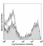
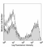
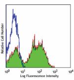
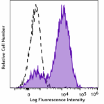
C57BL/6 mouse bone marrow cells were stained with biotinylated CD11b (clone M1/70) (filled histogram) or rat IgG2b, κ isotype control (open histogram), followed by Sav-PE (gated on total cells).
| Cat # | Size | Price | Quantity Check Availability | Save | ||
|---|---|---|---|---|---|---|
| 101203 | 50 µg | 34€ | ||||
| 101204 | 500 µg | 84€ | ||||
CD11b is a 170 kD glycoprotein also known as αM integrin, Mac-1 α subunit, Mol, CR3, and Ly-40. CD11b is a member of the integrin family, primarily expressed on granulocytes, monocytes/macrophages, dendritic cells, NK cells, and subsets of T and B cells. CD11b non-covalently associates with CD18 (β2 integrin) to form Mac-1. Mac-1 plays an important role in cell-cell interaction by binding its ligands ICAM-1 (CD54), ICAM-2 (CD102), ICAM-4 (CD242), iC3b, and fibrinogen.
Product DetailsProduct Details
- Verified Reactivity
- Mouse, Human, Cynomolgus, Rhesus
- Reported Reactivity
- Chimpanzee, Baboon, Rabbit
- Antibody Type
- Monoclonal
- Host Species
- Rat
- Immunogen
- C57BL/10 splenocytes
- Formulation
- Phosphate-buffered solution, pH 7.2, containing 0.09% sodium azide.
- Preparation
- The antibody was purified by affinity chromatography, and conjugated with biotin under optimal conditions.
- Concentration
- 0.5 mg/ml
- Storage & Handling
- The antibody solution should be stored undiluted between 2°C and 8°C. Do not freeze.
- Application
-
FC - Quality tested
- Recommended Usage
-
Each lot of this antibody is quality control tested by immunofluorescent staining with flow cytometric analysis. For flow cytometric staining, the suggested use of this reagent is ≤ 0.25 µg per 106 cells in 100 µL volume. It is recommended that the reagent be titrated for optimal performance for each application.
- Application Notes
-
Clone M1/70 has been verified for immunocytochemistry (ICC) and frozen immunohistochemistry (IHC-F).
Additional reported applications (for relevant formats of this clone) include: immunoprecipitation1,4, in vitro blocking3,9,12, depletion2,8, immunofluorescence microscopy6,7,10, immunohistochemistry of acetone-fixed frozen sections5,11-13, and spatial biology (IBEX)35,36. For in vivo studies or highly sensitive assays, we recommend Ultra-LEAF™ purified antibody (Endotoxin < 0.01 EU/µg, Azide-Free, 0.2 µm filtered) (Cat. No. 101248). -
Application References
(PubMed link indicates BioLegend citation) -
- Springer T, et al. 1978. Eur. J. Immunol. 8:539. (IP)
- Ault K and Springer T. 1981. J. Immunol. 126:359. (Deplete)
- Springer TA, et al. 1982. Immunol. Rev. 68:171. (Block)
- Ho MK and Springer TA. 1983. J. Biol. Chem. 258:2766. (IP)
- Flotte TJ, et al. 1983. Am. J. Pathol. 111:112. (IHC)
- Noel GJ, et al. 1990. J. Clin. Invest. 85:208. (IF)
- Allen LA and Aderem A. 1996. J. Exp. Med. 184:627 (IF)
- D'Amico A and Wu L. 2003. J. Exp. Med. 198:293. (Deplete)
- Brickson SJ, et al. 2003. Appl Physiol. 95:969. (Block)
- Clatworthy MR and Smith KG. 2004. J. Exp. Med. 199:717. (IF)
- Hata H, et al. 2004. J. Clin. Invest. 114:582. (IHC)
- Zhang Y, et al. 2002. J. Immunol. 168:3088. (IHC)
- Iwasaki A and Kelsall BL. 2001. J. Immunol. 166:4884 (IHC, FC)
- Tailleux L. 2003. J. Exp. Med. 197:121. (Block, FC)
- Olver S, et al. 2006. Cancer Research 66:571. (FC)
- Tan SL, et al. 2006. J. Immunol. 176:2872. (FC) PubMed
- Ponomarev ED, et al. 2006. J. Immunol. 176:1402. (FC)
- Dzhagalov I, et al. 2007. Blood 109:1620. (FC)
- Fazilleau N, et al. 2007. Nature Immunol. 8:753.
- Rasmussen JW, et al. 2006. Infect. Immun.74:6590. PubMed
- Napimoga MH, et al. 2008. J. Immunol. 180:609. PubMed
- Elqaraz-Carmon V, et al. 2008. J. Lipid. Res. 49:1894. PubMed
- Kim DD, et al. 2008. Blood 112:1109. PubMed
- Guo Y, et al. 2008. Blood 112:480. PubMed
- Norian LA, et al. 2009. Cancer Res. 69:3086. (FC) PubMed
- Baumgartner CK, et al. 2010. J. Immunol. 184:573. PubMed
- Charles N, et al. 2010. Nat. Med. 16:701. (FC) PubMed
- Whiteland J, et al. 1995. J. Histochem. Cytochem. 43:313. (IHC)
- Weber GF, et al. 2014. J Exp Med. 211:1243. PubMed
- Ashok A, et al. 2015. Toxicol Sci. 143:64. PubMed
- Price PJ, et al. 2015. J Immunol. 194:1164. PubMed
- Doni A, et al. 2015. J Exp Med. 212:905. PubMed
- Ferreira R, et al. 2016. J Infect Dis. 213: 669 - 673. PubMed
- Peterson VM, et al. 2017. Nat. Biotechnol. 35:936. (PG)
- Radtke AJ, et al. 2020. Proc Natl Acad Sci U S A. 117:33455-65. (SB) PubMed
- Radtke AJ, et al. 2022. Nat Protoc. 17:378-401. (SB) PubMed
- Product Citations
-
- RRID
-
AB_312786 (BioLegend Cat. No. 101203)
AB_312787 (BioLegend Cat. No. 101204)
Antigen Details
- Structure
- Integrin family, associates with integrin β2 (CD18), 170 kD
- Distribution
-
Granulocytes, monocytes/macrophages, dendritic cells, NK cells, subsets of T and B cells
- Function
- Adhesion, chemotaxis
- Ligand/Receptor
- ICAM-1 (CD54), ICAM-2 (CD102), ICAM-4 (CD242), iC3b, fibrinogen
- Cell Type
- B cells, Dendritic cells, Granulocytes, Macrophages, Monocytes, Neutrophils, NK cells, T cells, Tregs
- Biology Area
- Cell Adhesion, Cell Biology, Costimulatory Molecules, Immunology, Innate Immunity, Neuroscience, Neuroscience Cell Markers
- Molecular Family
- Adhesion Molecules, CD Molecules
- Antigen References
-
1. Barclay A, et al. 1997. The Leukocyte Antigen FactsBook Academic Press.
2. Springer TA. 1994. Cell 76:301.
3. Coxon A, et al. 1996. Immunity 5:653. - Gene ID
- 16409 View all products for this Gene ID 3684 View all products for this Gene ID
- UniProt
- View information about CD11b on UniProt.org
Related Pages & Pathways
Pathways


Related FAQs
- How many biotin molecules are per antibody structure?
- We don't routinely measure the number of biotins with our antibody products but the number of biotin molecules range from 3-6 molecules per antibody.
Other Formats
View All CD11b Reagents Request Custom ConjugationCustomers Also Purchased
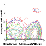
Compare Data Across All Formats
This data display is provided for general comparisons between formats.
Your actual data may vary due to variations in samples, target cells, instruments and their settings, staining conditions, and other factors.
If you need assistance with selecting the best format contact our expert technical support team.
-
PE/Cyanine7 anti-mouse/human CD11b
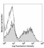
C57BL/6 mouse bone marrow cells were stained with CD11b (clo... -
APC/Fire™ 750 anti-mouse/human CD11b
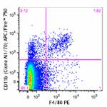
C57BL/6 splenocytes were blocked with TruStain fcX™ (a... 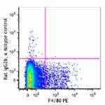
-
TotalSeq™-A0014 anti-mouse/human CD11b
-
Brilliant Violet 750™ anti-mouse/human CD11b
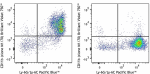
C57BL/6 mouse bone marrow cells were stained with Ly-6G/Ly-6... -
TotalSeq™-C0014 anti-mouse/human CD11b
-
TotalSeq™-B0014 anti-mouse/human CD11b
-
Spark NIR™ 685 anti-mouse/human CD11b
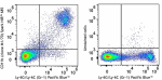
C57BL/6 splenocytes were stained with Ly-6G/Ly-6C (Gr-1) Pac... -
PE/Fire™ 640 anti-mouse/human CD11b
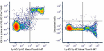
C57BL/6 mouse bone marrow cells were stained with anti-mouse... -
APC anti-mouse/human CD11b
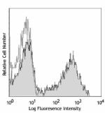
C57BL/6 mouse bone marrow cells were stained with CD11b (clo... -
Biotin anti-mouse/human CD11b
C57BL/6 mouse bone marrow cells were stained with biotinylat... -
FITC anti-mouse/human CD11b
C57BL/6 mouse bone marrow cells were stained with CD11b (clo... 
-
PE anti-mouse/human CD11b
C57BL/6 mouse bone marrow cells were stained with CD11b (clo... 
Fixed whole mount mouse spleen was stained with Alexa Fluor®... -
PE/Cyanine5 anti-mouse/human CD11b
C57BL/6 mouse bone marrow cells stained with CD11b (clone M1... -
Purified anti-mouse/human CD11b
C57BL/6 mouse bone marrow cells were stained with purified C... 
Fresh, frozen mouse spleen was stained with purified CD11b c... -
Alexa Fluor® 488 anti-mouse/human CD11b
C57BL/6 mouse bone marrow cells were stained with CD11b (clo... 
C57BL/6 mouse frozen spleen section was fixed with 4% parafo... 
Paraformaldehyde-fixed (1%), 500 µm-thick mouse spleen secti... 
Confocal image of C57BL/6 mouse liver sample acquired using ... -
Alexa Fluor® 647 anti-mouse/human CD11b
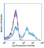
C57BL/6 mouse bone marrow cells were stained with CD11b (clo... 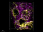
Paraformaldehyde-fixed (4%), 500 µm-thick mouse spleen secti... -
Alexa Fluor® 700 anti-mouse/human CD11b
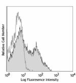
C57BL/6 mouse bone marrow cells were stained with CD11b (clo... -
Pacific Blue™ anti-mouse/human CD11b
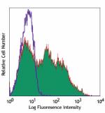
C57BL/6 mouse bone marrow cells were stained with CD11b (clo... -
APC/Cyanine7 anti-mouse/human CD11b
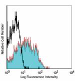
C57BL/6 mouse bone marrow stained with M1/70 APC/Cyanine7 -
PerCP/Cyanine5.5 anti-mouse/human CD11b
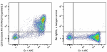
C57BL/6 splenocytes were blocked with TruStain FcX™ (a... -
PerCP anti-mouse/human CD11b
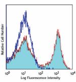
C57BL/6 mouse bone marrow cells were stained with CD11b (clo... -
Brilliant Violet 421™ anti-mouse/human CD11b
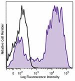
C57BL/6 mouse bone marrow cells were stained with CD11b (clo... 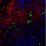
BL/6 mouse lymph nodes, fixed O/N in PLP, blocked with 10% r... -
Brilliant Violet 570™ anti-mouse/human CD11b
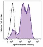
C57BL/6 mouse bone marrow cells were stained with CD11b (clo... -
Brilliant Violet 605™ anti-mouse/human CD11b

C57BL/6 mouse bone marrow cells were stained with CD11b (clo... -
Brilliant Violet 650™ anti-mouse/human CD11b
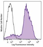
C57BL/6 mouse bone marrow cells were stained with CD11b (clo... -
Brilliant Violet 711™ anti-mouse/human CD11b
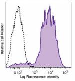
C57BL/6 mouse bone marrow cells were stained with CD11b (clo... -
Brilliant Violet 785™ anti-mouse/human CD11b
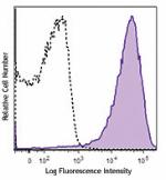
C57BL/6 mouse bone marrow cells were stained with CD11b (clo... -
Brilliant Violet 510™ anti-mouse/human CD11b
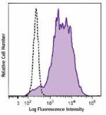
C57BL/6 mouse bone marrow cells were stained with CD11b (clo... 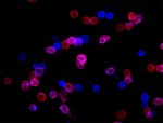
C57 mouse bone marrow cells were fixed with 2% paraformaldeh... -
Ultra-LEAF™ Purified anti-mouse/human CD11b
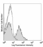
C57BL/6 mouse bone marrow cells were stained with Ultra-LEAF... -
Purified anti-mouse/human CD11b (Maxpar® Ready)
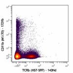
Mouse splenocytes stained with 172Yb-anti-CD11b (M1/70) and ... -
Alexa Fluor® 594 anti-mouse/human CD11b
C57BL/6 mouse frozen spleen section was fixed with 4% parafo... 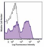
C57BL/6 mouse bone marrow cells were stained with CD11b (clo... -
PE/Dazzle™ 594 anti-mouse/human CD11b
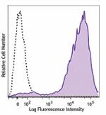
C57BL/6 mouse bone marrow cells were stained with CD11b (clo... -
Spark YG™ 593 anti-mouse/human CD11b
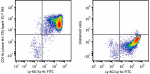
C57BL/6 mouse bone marrow cells were stained with Ly-6G/Ly-6... -
Spark YG™ 570 anti-mouse/human CD11b
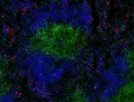
C57BL/6 mouse frozen spleen section was fixed with 4% parafo... -
PE/Fire™ 810 anti-mouse/human CD11b
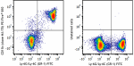
C57BL/6 mouse bone marrow cells were stained with anti-mouse... -
APC/Fire™ 810 anti-mouse/human CD11b Antibody
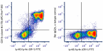
C57BL/6 mouse bone marrow cells were stained with anti-mouse... -
Spark Blue™ 550 anti-mouse/human CD11b
C57BL/6 mouse bone marrow cells were stained with anti-mouse... -
Spark UV™ 387 anti-mouse/human CD11b
C57BL/6 bone marrow cells were stained with anti-mouse Ly-6G... -
PerCP/Fire™ 806 anti-mouse/human CD11b
C57BL/6 mouse bone marrow cells were stained with anti-mouse... -
PerCP/Fire™ 780 anti-mouse/human CD11b
C57BL/6 mouse bone marrow cells were stained with anti-mouse... -
Spark Blue™ 574 anti-mouse/human CD11b (Flexi-Fluor™)
-
KIRAVIA Blue 520™ anti-mouse/human CD11b
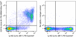
C57BL/6 mouse bone marrow cells were stained with anti-mouse... -
PE/Fire™ 744 anti-mouse/human CD11b

C57BL/6 mouse bone marrow cells were stained with anti-mouse... -
Spark PLUS UV395™ anti-mouse/human CD11b

C57BL/6 mouse splenocytes were stained with anti-mouse Ly-6G... -
Spark Red™ 718 anti-mouse/human CD11b (Flexi-Fluor™)
-
Spark PLUS B550™ anti-mouse/human CD11b

C57BL/6 mouse bone marrow cells were stained with anti-mouse... -
Spark YG™ 581 anti-mouse/human CD11b (Flexi-Fluor™)
-
StarBright UltraViolet 575 anti-mouse/human CD11b

C57BL/6 mouse bone marrow cells were stained with anti-mouse... -
StarBright UltraViolet 740 anti-mouse/human CD11b

C57BL/6 mouse bone marrow cells were stained with anti-mouse... -
Spark PLUS V475™ anti-mouse/human CD11b

C57BL/6 mouse bone marrow cells were stained with anti-mouse... -
Spark PLUS YG581™ anti-mouse/human CD11b

C57BL/6 mouse bone marrow cells were stained with anti-mouse... -
Spark PLUS V450™ anti-mouse/human CD11b

C57BL/6 mouse bone marrow cells were stained with anti-mouse...


Follow Us